Pathologies

In our specialized center we treat all eye pathologies, as we have super-specialists in all subspecialties of modern ophthalmology.
Myopia:
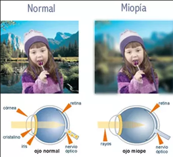
It is an optical problem that prevents seeing distant objects clearly. This is because the myopic eye has a greater axial length than normal and/or a more curved cornea than normal. Then the rays that enter the eye focus in front of the retina producing blurred vision of distant objects. These people can see near objects well, but need prescription lenses.
View Brochure

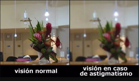
Astigmatism: It is the optical problem caused by an uneven curvature in the cornea or lens, with one meridian more curved and another flatter. When parallel rays enter the eye, they focus in more than one position, causing distorted and blurred images. To improve Astigmatism, cylindrical lenses are prescribed with their axis oriented at 90º from the meridian to be corrected.
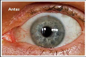
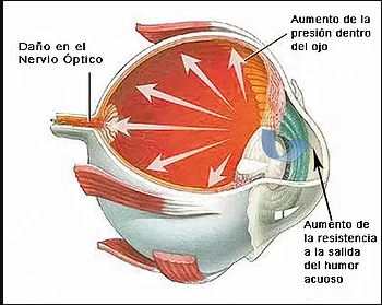
Glaucoma: glaucoma_desc_1
glaucoma_desc_2
View Brochure
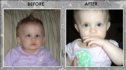
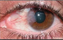
Pterygium: The conjunctiva invades the cornea, it is a tip of conjunctival tissue that advances and grows from the outer part of the cornea towards the center.
They are cells and blood vessels from the conjunctiva that enter, invade and place themselves in front of the cornea, potentially obstructing vision if they reach the pupil.
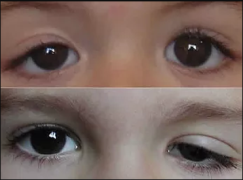
Ptosis:
Palpebral ptosis, also called blepharoptosis or eyelid ptosis, is a permanent descent of the upper eyelid. It can be total or partial, depending on whether it prevents vision or not. This symptom can be caused by eyelid damage (the term Blepharoptosis is then used) or by nerve damage of the third pair or brain nerve centers (in this case, the word ptosis is frequently used).
View Brochure
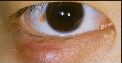
Stye: A stye often appears as an irritated bump near the edge of the eyelid, caused by an infected eyelash follicle.
It is important not to squeeze or try to remove a stye or chalazion.
View Brochure
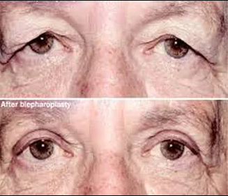
Dermatochalasis: It is a process associated with aging or pseudo-herniation of fat bags characterized by flaccidity of the upper eyelid skin, producing redundancy that can reach the margin of the eyelashes.
It originates from the loss of elasticity and stretching of the upper eyelid skin that occurs with age.
View Brochure

Hyperopia: It is an optical problem opposite to myopia, in which the patient's eye has a shorter axial length than normal and/or a very flat cornea. For this reason, the rays focus behind the retina, causing nearby objects to appear distorted, while distant ones are seen normally. To see clearly, these people must use positive graduation lenses.
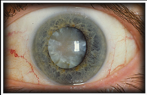
Cataracts: A cataract is the total or partial opacification of the lens. The opacification causes light to scatter inside the eye and cannot be focused on the retina, creating diffuse images.
It is the most common cause of blindness treatable with surgery. It has various causes but is mostly attributed to age although there are many other causes.
View Brochure
Strabismus:
Strabismus is the deviation of the alignment of one eye in relation to the other, preventing bifoveolar fixation. This prevents fixing the gaze of both eyes at the same point in space, which causes incorrect binocular vision that can adversely affect depth perception.
View Brochure
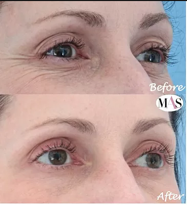
Wrinkles, Folds, and Crow's Feet: Wrinkles appear as a result of aging processes. They form as a consequence of: decreased collagen, genetic factors, facial expressions, sun damage, poor hydration, or smoking.
To treat them, Botox is implemented, which acts by relaxing that musculature with a localized and natural action preventing the formation of wrinkles. The therapeutic effect of Botox varies in each patient, averaging 6 to 8 months.
View Brochure
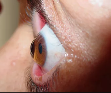
Keratoconus: It is an eye disease that affects the structure of the cornea, the transparent tissue that covers the front of the eye.
The shape of the cornea slowly changes from the normal round shape to a conical shape. Then when light enters the eye through the cornea, it does not focus the light rays correctly causing visual problems.
View Brochure
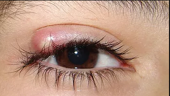
Chalazion:
A chalazion forms when a sebaceous gland in the eyelid called the Meibomian gland enlarges and the gland opening becomes blocked due to fat.
View Brochure
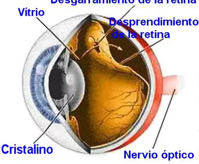
The retina is the tissue in the back of the eye that helps you see the images focused on it by the cornea and lens.
When retinal detachment occurs, bleeding from nearby blood vessels can cause opacity inside the eye, so that you may not see clearly or not see at all. Central vision can be seriously affected if the macula, the part of the retina responsible for fine and detailed vision, becomes detached.
View Brochure